Overcoming the limits of optical resolution and increasing numerical aperture (NA) are two of the main problems in light microscopy.
I will cover both resolution and NA in other articles, but in brief, the NA of an objective relates to the light gathering abilities whereas resolution relates to the ability distinguish details in a specimen. In this article, we will have a look at microscopy immersion techniques for imaging specimens at high magnifications whilst increasing NA and overcoming some of the limits of resolution.
If the air gap between the objective front lens and the cover glass of a slide is replaced with an immersion liquid, then we are able to somewhat increase the resolution of an objective which, in turn, will increase the NA. As light passes from one material or medium to another (for example, from air to glass) it undergoes ‘refraction’- which simply means that it scatters and bends. Light rays from a specimen can be refracted when travelling from the glass of the coverslip into the air, or may be reflected by the coverslip or physically blocked by the housing surrounding the objective front lens. Each of these aberrations will not contribute to the final image formation. If we replace the air gap between the coverslip and the objective front lens, then we can somewhat decrease the refraction (and reflection) of light coming from the specimen and consequently increase the ability of the objective to capture this otherwise deviated light.
The refractive index
The amount of refraction is determined by the physical properties of the medium through which light rays pass. This ‘amount of refraction’ is described as the ‘refractive index’. This is a numerical value (without units) which describes the extent to which light refracts when passing through a medium or material. For example, air has a refractive index of 1.0 whereas microscope slides (and coverslips) have refractive indexes of 1.5. The purpose of any immersion liquid is to closely match the refractive index of the glass slides, which consequently increases the amount of light which will form the final specimen image. Most commercial immersion oils have a refractive index of 1.51 to match the index of the glass.
Working distance
Another factor of microscope objectives to consider is the ‘Working Distance’. When a specimen is in sharp focus, the distance between the front lens of an objective and the surface of the coverslip is known as the ‘working distance’. As an objective is moved closer to the specimen and slide, the focal plane will subsequently move further into the specimen. Obviously, an objective can only be moved until it is in contact with a coverslip. As the magnification of an objective increases, the working distance will decrease. For example, a 10X objective will have an approximate working distance of 4 mm, conversely, the working distance of a 100X oil immersion objective will be around 130 microns. The working distance of an objective will usually be found on the objective barrel (abbreviated as ‘WD’).
Immersion oils
Oil of cedar wood was routinely used for immersion microscopy for many years (and is still available today). Despite having a refractive index of 1.516, this oil can turn yellow with age and could damage the objective front lens if not removed immediately after use. Furthermore, In addition, cedar wood oil absorbs blue and ultraviolet light.
When using immersion oils, you should always use correctly matched immersion oils which are recommended by the objective manufacturer.
Many modern commercial oils are synthetically manufactured and standardised to avoid damage to lenses and remain colour-stable over time. One thing to bear in mind is that immersion oils have optimal working temperatures. Most of the synthetic oils are designed to work at the average room temperature of 230C. A change in temperature of only 10C will increase or decrease the refractive index of the immersion oil by a factor of 0.0004. This factor should be taken into account if, for example, you are planning long-term live-cell imaging experiments during which the environment immediately surrounding the cells on the microscope stage will be at 370C. Consequently, oils are available which are designed for such temperatures.
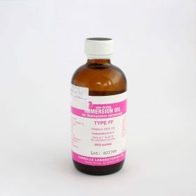
You can find a variety of immersion oils available from Agar Scientific including Cargille oils;
http://www.agarscientific.com/cargille-immersion-oils.html
Finally, you should also be aware that many of the general immersion oils will auto-fluoresce. If you are carrying out fluorescence microscopy experiments, then you should use one of the immersion oils designed to work under fluorescent conditions. Such oils have the letter ‘F’ before or after the oil name or code.
Practical tips for using oil immersion
- Firstly, image your specimen using a low power objective to find the area of interest on your slide.
- Work your way from the low power objective to the 40X objective whilst keeping your area of interest in view. Then set up the microscope for Koehler Illumination (see my article on this subject which explains the correct set-up).
- Swing the objective turret (‘nosepiece’) around between the 40X and the oil immersion objective without engaging the high magnification objective.
- Look away from the eyepieces to the side of the microscope. Place one drop of immersion oil directly on to the coverslip (that’s ‘one drop’- you are working with a sensitive optical instrument, not oiling a bike chain!). Carefully turn the nosepiece to fully engage the high power objective. Whilst continuing to look from the side of the microscope, slowly use the focus wheels to bring the objective front lens into contact with the immersion oil drop on the coverslip. If you are using an oil objective which has a concave front lens then you should also add a drop of immersion oil to this lens to prevent any air bubbles becoming trapped between the oil drop and the concave recess.
- Look down the eyepieces once again. Using only the fine focus from now on, slowly bring your specimen into focus. Most oil immersion objectives have spring-loaded nose cones, but rapid or coarse focussing at this stage could easily crack the coverslip and slide resulting in shards of glass and oil falling into the substage condenser (on an upright microscope) or over the objectives and nosecone (on an inverted microscope). Such an accident may also damage the objective front lens, so avoid this at all cost!
- Once you have finished using the oil immersion objective, clean the oil from the objective, even if you are planning to have a look at other slides. This will prevent possible contamination of other objectives or components. Immersion oil can damage other parts of the microscope components especially objectives which are not designed for this purpose. Immersion oil can corrode the cement which holds objective front lenses in place. To remove the oil, use a lens cleaning tissue in a single sweeping motion across the objective front lens. Continue to wipe with a clean piece of tissue for each sweep until no trace of oil remains on the tissue. A small amount of xylene can be used for final cleaning or commercial oil removal solutions are available. As before, you should check the manufacturer’s recommendations before using any solutions on the objectives.
AUTHOR: Martin Wilson


